How to analyze samples by custom-made (self-made) microarrays.
Flexible experimental systems, not available with conventional products
1.Overview
This technical note describes an assay method using the substrate for PepTenChip®. Biochips provide various applications in the laboratory level, such as simple detection, monitoring, and screening of proteins and/or toxins contained in samples. This technical note uses a substrate immobilized with biotin as the capture molecule and employs a solution containing dye (TAMRA as a example)-labeled streptavidin as an analyte. Fluorescence measurements were performed by fluorescence detector (PepTenCam).
A demonstration video is also available online.
2.Assay Procedure
This procedure describes how to apply the sample solution to the substrate, incubate it, and then measure it using a fluorescence detector. For substrate derivatization and array procedures, refer to the other technical note “Substrate Derivatization and Manual Arraying (TO14E)”.
2-1 Blank Measurement
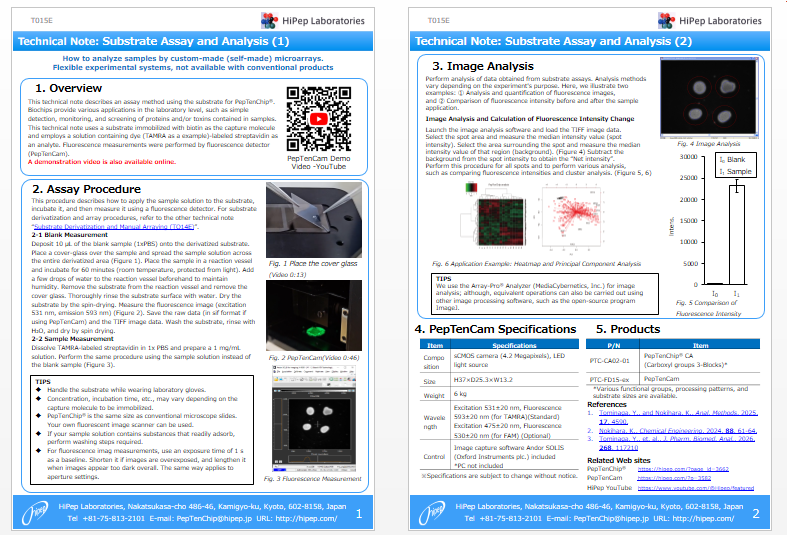
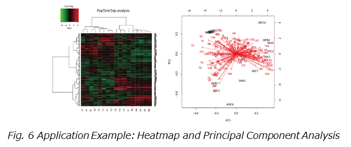
Deposit 10 µL of the blank sample (1xPBS) onto the derivatized substrate. Place a cover-glass over the sample and spread the sample solution across the entire derivatized area (Figure 1). Place the sample in a reaction vessel and incubate for 60 minutes (room temperature, protected from light). Add a few drops of water to the reaction vessel beforehand to maintain humidity. Remove the substrate from the reaction vessel and remove the cover glass. Thoroughly rinse the substrate surface with water. Dry the substrate by the spin-drying. Measure the fluorescence image (excitation 531 nm, emission 593 nm) (Figure 2). Save the raw data (in sif format if using PepTenCam) and the TIFF image data. Wash the substrate, rinse with H₂O, and dry by spin drying.
2-2 Sample Measurement
Dissolve TAMRA-labeled streptavidin in 1x PBS and prepare a 1 mg/mL solution. Perform the same procedure using the sample solution instead of the blank sample (Figure 3).
TIPS
- Handle the substrate while wearing laboratory gloves.
- Concentration, incubation time, etc., may vary depending on the capture molecule to be immobilized.
- PepTenChip® is the same size as conventional microscope slides. Your own fluorescent image scanner can be used.
- If your sample solution contains substances that readily adsorb, perform washing steps required.
- For fluorescence imag measurements, use an exposure time of 1 s as a baseline. Shorten it if images are overexposed, and lengthen it when images appear too dark overall. The same way applies to aperture settings.
3.Image Analysis
Perform analysis of data obtained from substrate assays. Analysis methods vary depending on the experiment’s purpose. Here, we illustrate two examples: ① Analysis and quantification of fluorescence images,
and ② Comparison of fluorescence intensity before and after the sample application.
Image Analysis and Calculation of Fluorescence Intensity Change
Launch the image analysis software and load the TIFF image data.
Select the spot area and measure the median intensity value (spot intensity). Select the area surrounding the spot and measure the median intensity value of that region (background). (Figure 4) Subtract the background from the spot intensity to obtain the “Net intensity”.
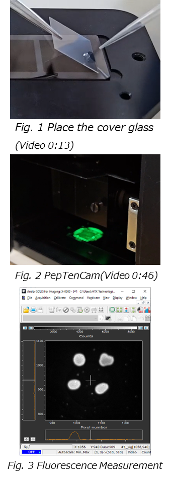
Perform this procedure for all spots and to perform various analysis,
such as comparing fluorescence intensities and cluster analysis. (Figure 5, 6)


TIPS
We use the Array-Pro® Analyzer (MediaCybernetics, Inc.) for image analysis; although, equivalent operations can also be carried out using other image processing software, such as the open-source program ImageJ.
4.PepTenCamSpecifications
| Item | Specifications |
| Composition | sCMOS camera (4.2 Megapixels), LED light source |
| Size | H37×D25.3×W13.2 |
| Weight | 6 kg |
| Wavelength | Excitation 531±20 nm, Fluorescence 593±20 nm (for TAMRA)(Standard) Excitation 475±20 nm, Fluorescence 530±20 nm (for FAM) (Optional) |
| Control | Image capture software Andor SOLIS (Oxford Instruments plc.) included *PC not included |
5.Products
| P/N | Item |
| PTC-CA02-01 | PepTenChip® CA (Carboxyl groups 3-Blocks)* |
| PTC-FD15-ex | PepTenCam |
References
1.Tominaga, Y., and Nokihara, K., Anal. Methods, 2025, 17, 4590.
2.Nokihara, K., Chemical Engineering, 2024, 88, 61-64.
3.Tominaga, Y., et. al., J. Pharm. Biomed. Anal., 2026, 268, 117210
Related Web sites
PepTenChip® https://hipep.com/?page_id=3662
PepTenCam https://hipep.com/?p=3582
HiPep YouTube https://www.youtube.com/@Hipep/featured