
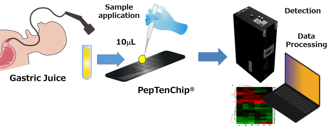
Overview
This product is the research-grade microarray (biochip), featuring 40 selected capturing peptides (discovered by HiPep) focusing on precancerous gastric lesions (such as atrophic gastritis and intestinal metaplasia) arrayed on amorphous carbon substrates. Minute samples of gastric fluid are required to assess the risk of progression to gastric cancer without pathological tissue biopsies. PepTenCam is a suitable detector, although measurements can also be performed with your in-house fluorescence detectors and/or scanners.
Features
■Arrayed with selected capture molecules, which enable highly accurate stratification of precancerous gastric lesions: selected peptides have high correlation in identifying precancerous conditions and the progression (risk stratification) of early-stage gastric lesions.
■Amorphous carbon substrates allow ultra-high sensitivity and low-noise detection. The substrate provides minimized non-specific adsorption (background noise) to the utmost extent, enabling signal detection with a high signal-to-noise ratio. Additionally, PepTenChip® is not disposable but reusable, offering long-term cost-effectiveness with environmentally beneficial.
■Comprehensive and rapid multiplex analysis: Since multiple biomolecules in analytes can be evaluated simultaneously on a single chip, sample consumption and analysis time are significantly reduced.
| P/N Product | Unit | Price |
| PTC-GCM-01 Stomach Cancer Risk (Pre-disease) classification Chip | 1 | Contact us |
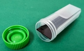
| Tip storage case | 1 | Included |
| Incubation reaction vessels | 1 | Included |
Gastric Cancer Risk (Pre-disease) Classification Chip
| Capture Peptides | 40 types of α-Helical peptides (Seq. α-TAMRA-G-XXXX-GC-NH2) |
| Substrate | Maleimide-coated amorphous carbon substrate with 3 Block design |
| Immobilization Method | Immobilized via a covalent bond between the SH group of the peptide-size cysteine residue and the polymethylene linker on the substrate surface through a maleimide group |
| Spot diameter | ca 160 μm |
| Pitch | ca 450 μm |
| Array method | ca 0.14 pmol of peptide was arrayed per spot by microarray spotter |
| Storage | Store in its dedicated case, keep in dark at 4°C |
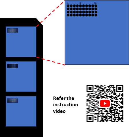
Array Map (Location) and List of Peptides
| Spot | Probe# | Seq.(XXXX) |
| 1 | A0105 | LRRLLSLLRRLLSL |
| 2 | A0109 | LSSLLRALRRLLSA |
| 3 | A0110 | LSSLLRLLRRLLSL |
| 4 | A0115 | LKKLIKILKKLIKI |
| 5 | A0120 | LKKLIRILKKLIRI |
| 6 | A0121 | LKKLLRILKKLLRI |
| 7 | A0122 | LRRLIKILKKLIRI |
| 8 | A0123 | LRRLLKILKKLLRI |
| 9 | A0124 | LRRLIKILRRLIKI |
| 10 | A0125 | LRRLLKILRRLLKI |
| 11 | A0126 | LKKLIEILKKLIEI |
| 12 | A0127 | LKKLLEILKKLLEI |
| Spot | Probe# | Seq.(XXXX) |
| 13 | A0129 | LEELLKILKKLLEI |
| 14 | A0138 | LKKLISILKKLISI |
| 15 | A0139 | LKKLLSILKKLLSI |
| 16 | A0140 | LSSLIKILKKLISI |
| 17 | A0141 | LSSLLKILKKLLSI |
| 18 | A0144 | LRRLIEILRRLIEI |
| 19 | A0157 | LRRLLSILRRLLSI |
| 20 | A0165 | LKKLLKFLKKLLKF |
| 21 | A0173 | LKKLLRFLKKLLRF |
| 22 | A0185 | LKKLLEFLKKLLEF |
| 23 | A0213 | LSSLLKFLKKLLSF |
| 24 | A0254 | LKKLVKVLKKLVKV |
| Spot | Probe# | Seq.(XXXX) |
| 25 | A0255 | LKKLLKVLKKLLKV |
| 26 | A0258 | LKKLVRVLKKLVRV |
| 27 | A0259 | LKKLLRVLKKLLRV |
| 28 | A0260 | LRRLVKVLKKLVRV |
| 29 | A0261 | LRRLLKVLKKLLRV |
| 30 | A0265 | LKKLLEVLKKLLEV |
| 31 | A0267 | LEELLKVLKKLLEV |
| 32 | A0269 | LEELLKVLEELLKV |
| 33 | A0272 | LQQLVKVLKKLVQV |
| 34 | A0275 | LQQLLKVLQQLLKV |
| 35 | A0277 | LKKLLSVLKKLLSV |
| 36 | A0279 | LSSLLKVLKKLLSV |
| 37 | A0291 | LQQLLRVLRRLLQV |
| 38 | A0420 | AKKAARVAKKAARV |
| 39 | A0457 | ASSAVRVARRAVSV |
| 40 | A0483 | IKKFFSFIKKFFSF |
Operation Manual
1. Preparation and Setup:
Place the substrate on a level, clean surface free of dust and debris. Measurement of fluorescence intensity of the substrate, I0 data using PBS before sample application.
2. Sample Application and Incubation (for 3 blocks) *For each block, perform steps ① and ② in the following order.
① Sample Application: Accurately dispense 10 μL of sample onto each block.
② Cover Glass Placement: Immediately and gently place the cover glass on top to ensure the
dispensed sample spreads evenly across the entire block.
③Processing the Remaining Blocks: Quickly repeat steps ① and ② for the remaining two blocks.
3. Incubation and Measurement
① Sealing: Immediately place the processed plate into the “Plate Reaction Vessel,” close the cap
securely, and seal it.
② Incubation: Leave the plate at room temperature in a dark place for 15 to 30 minutes.
③ Fluorescence Measurement: After incubation, remove the plate and perform the measurement
using a fluorescence detector, I1.
💡 Key Points for Accurate Measurement
Preventing air bubbles: When placing the cover glass, tilt it gently from the edge to avoid trapping air bubbles, which cause noise and/or signal loss during detection.
Avoid drying: Keep the time between dispensing the sample and placing the cover glass as short as possible.
Thorough Light Shielding: Important to store the chip in “dark” to prevent the fluorescent substance from fading. It is recommended that wrapping the container with aluminum foil or storing it in a light-shielding box.
Related Products:
Fluorescence Detector
PepTenCam (PTC-FD15, CP06E)
Other TESTING chip series for research-use:
★ Oral healthcare rapid detection chip:
P/N PTC-PDS-01
★ Cerebrospinal fluid Classification Chip
(focusing on multiple sclerosis and related diseases):
P/N PTC-NDS-01 (Coming soon)
Reference
[1] Nokihara, K., Chemical Engineering, 2024, 88, 61-64.
[2] Tominaga, Y., et. al. (2018) Bioorg. Med. Chem., 26, 3210-3216.
[3] Tominaga, Y. and Nokihara , K. (2025) Anal. Meth. (Royal Society of Chemistry), 17, 4590-4598.
[4] Tominaga, Y., Wu, X., Wei, M., and Nokihara. K. (2026) J. Pharm. Biomed. Anal. 268,117210.
Videos: Summary Video on Bio-Detection Methods Based on New Principles https://hipep.com/?page_id=3662
①PepTenChip®/PepTenCam Videos https://youtu.be/-Zli6QZVetU
②Introducing New Carbon Substrate Material https://www.youtube.com/watch?v=xcar8LTKAcU
③Manual Array Preparation Method: Researchers without an arrayer can easily create arrays.
You can create arrays using your own molecules. Protocol for Reproduction and Reuse https://www.youtube.com/watch?v=bFVfJTDY4Uw